QUICK FACTS
Milestone: Autopsy on famous patient “Tan”
Date: April 18, 1861
Where: Bicêtre Hospital, outside Paris
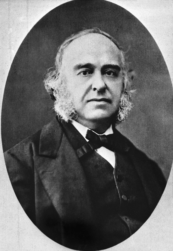
Who: Dr. Paul Broca
On April 18, 1861, a doctor in Paris cut open the brain of a patient who had died the day before — and unwittingly identified a brain region that’s key to spoken language.
The patient, Louis Victor Leborgne, was nicknamed “Tan” by doctors at Bicêtre Hospital because it was one of the only words he could say. By the time he died at age 51, he had spent 21 years living in the psychiatric ward of the hospital.
Leborgne was reportedly healthy at birth but began having epileptic seizures in early childhood. At age 30, he lost his ability to speak. For a while, he avoided getting treatment, but he was eventually admitted to Bicêtre Hospital.
Article continues below
Doctors found that he could understand language well and he would use gestures to convey his needs. Rarely, he could utter a swear word.
A decade after he was admitted to the hospital, he began to experience right-sided paralysis that grew steadily worse, as well as mental difficulties. Eventually, he lost the ability to walk. He spent the last seven years of his life in bed.
During these last few years, Dr. Paul Broca, a surgeon at the hospital, began to see Leborgne as a patient.
“The numerical responses were the ones he made best, by opening or closing his fingers. He would indicate, without error, the time on a watch to the second. He knew exactly how many years he had been in Bicêtre, etc,” Broca said of his patient, according to a translation.
“However, many questions to which a man of normal intelligence would have found the means to respond by gesture, remained without intelligible response; other times the response was clear, but did not answer the question,” Broca observed. “Undoubtedly, then, the intelligence of the patient had been affected to a great degree, but he maintained certainly more of it than was needed for talking.”
On April 17, 1861, Leborgne died of gangrene — likely a result of a bedsore in his leg. The next day, Broca began an autopsy and noted a pocket of clear fluid about the size of a “chicken’s egg” in the perisylvian region of the brain’s left hemisphere; this region surrounds a deep groove called the lateral sulcus, which marks the upper boundary of the temporal lobe. Several areas surrounding the fluid exhibited a “softness.” And there were other abnormalities: Leborgne’s brain was lighter than normal, and several brain regions had a smaller volume than expected.
That same day, Broca presented his autopsy findings at the Anthropological Society Meeting in Paris. At the time, there was an ongoing debate between scientists who believed all of the brain’s functions were diffused throughout the organ’s tissues and those who believed certain regions performed specific functions.
Broca’s autopsy was strong evidence for the latter idea.
“The principal home and the original seat of the softness is the middle part of the frontal lobe of the left hemisphere; it is there that one finds the most extensive lesions — the most advanced and the oldest,” he said in his presentation.
This suggested that “in the present case, the lesion of the frontal lobe was the cause of the loss of speech,” Broca added.
At the meeting, however, his peers didn’t immediately recognize the finding’s significance; most of the meeting was taken up with now-discredited race “science” focused on supposed links between skull measurements and intelligence. But by August 1861, Broca had studied the brains of multiple patients with what would later be termed aphasia. The research reinforced his conviction that speech was localized to the frontal lobe, and he would later narrow the region to the left frontal lobe.
Over the course of his life, Broca would not only identify the region tied to aphasia but also note that speech therapy could occasionally help patients regain speech.
Since Broca’s time, researchers have confirmed that discrete brain regions perform specific cognitive functions and have zeroed in on a much more precise region of the brain that is key for speech than Broca identified. That area is now named Broca’s area and is recognized as important to Broca’s aphasia, in which patients can understand language but have trouble producing spoken, written or sign language.
We now know that other regions and networks beyond Broca’s area play a big role in speech. For instance, damage to Wernicke’s area, discovered in 1874, can trigger a form of aphasia in which patients speak in long, complete sentences that have little meaning.
For decades, Leborgne’s intact brain, which Broca never cut into sections but only examined superficially, could be viewed at the Dupuytren Museum in Paris, which closed to the public in 2016.
Source: Read Full Article